| Home | Accessory Kit | Marsh CD Collection | Library | Contact Us |
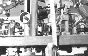
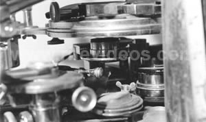
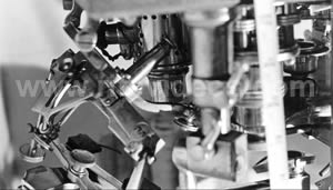
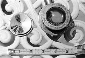
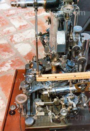
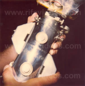
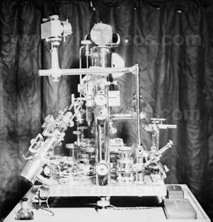
The Universal Microscope |
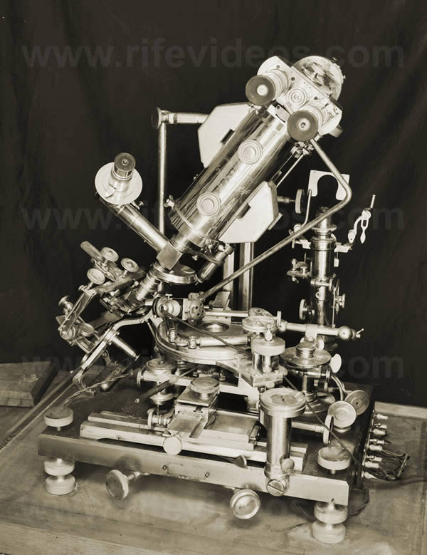 |
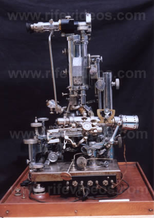
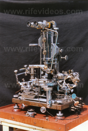
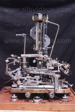
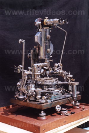
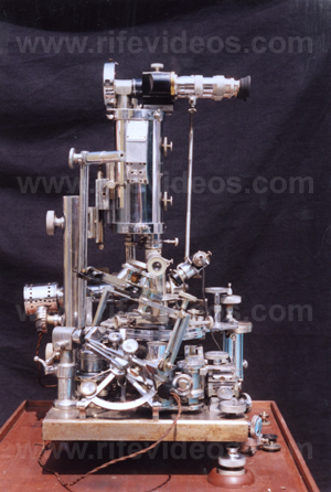
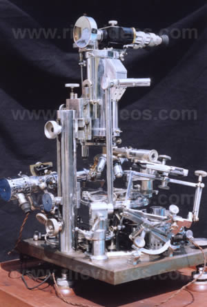
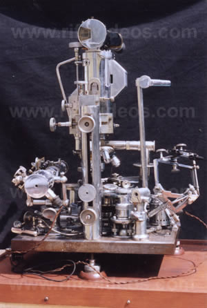
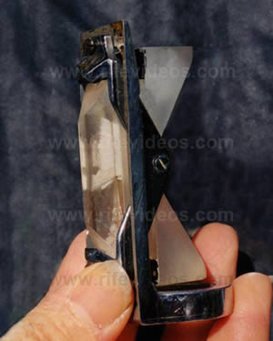
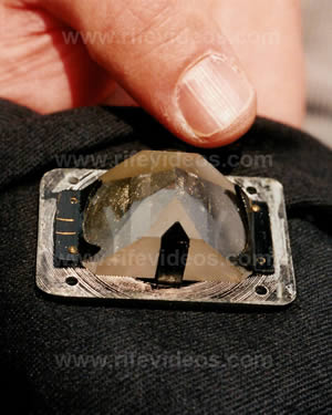
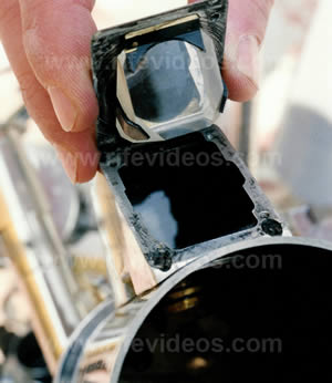
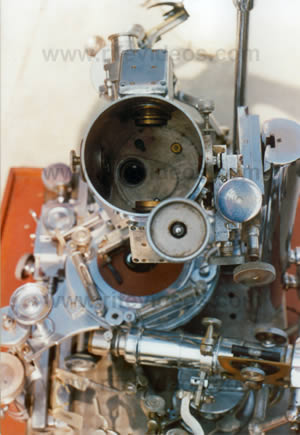
1. Crane: Dr. Rife. 2. Crane: This is John Crane now breaking in on the conversation with Dr. Rife to go back and review some of the fundamentals of the universal microscope. The universal microscope was designed by Dr. Royal R. Rife who for many years has built and worked with light microscopes which surpassed the limitations of the best standard microscopes. All the Rife microscopes possess superior abilities to attain higher resolution and magnification. The largest and most powerful of these is the Universal Microscope, built in 1933, consisting of 5,280 parts and is so called because of its adaptability in all fields of microscopical work, being fully equipped with separate substage units of the condenser for transmitted and monochromatic beam dark-field, polarized, and slit-ultra illumination, including also a special system for crystallography work. The entire optical system of lenses and prisms as well as the illuminating units are made of block-crystal quartz, quartz being especially transparent to ultraviolet radiations. The illuminating unit used for examining the filterable forms of disease organisms contains 14 lenses and prisms, 3 of which are in the high-intensity incandescent lamp, 4 in the Risley prism, and 7 in the achromatic condenser which has a numerical aperture of 1.40. Between the source of light and the specimen are subtended two circular, wedge-shaped, block-crystal quartz prisms for the purpose of polarizing the light passing through the specimen, polarization being the practical application of the theory that light waves vibrate in all planes perpendicular to the direction in which they are propagated. Therefore, when light comes into contact with a polarizing prism, it is divided or split into two beams… Rife: Wait a minute John, I want to hold that up because this is not true polarized light, this is the monochromatic beam illumination, which is their error (1944 Smithsonian Report) when they copied that off. This is their error, and it is a monochromatic beam which is the separation of a certain portion of a spectrum, see, go ahead… 3.Crane: Therefore, when light comes into contact with a polarizing prism, it is divided or split into two beams one of which is refracted to such an extent that it is reflected to the side of the prism without passing through the prism. While the second ray, bent considerably less, is thus enabled to pass through the prism to illuminate the specimen. This is the portion of the monochromatic beam, I presume? Rife: Yes, that’s it, yes. 4. Crane: When the quartz prisms on the universal microscope may be rotated. When they are rotated through 360 degrees they are rotated in opposite directions. Then they serve to bend the transmitted light at variable angles of incidence while, at the same time, a spectrum is projected up into the axis of the microscope, or rather a small portion of the spectrum. Since only a part of the color band is visible at any time it is possible to proceed in this way from one end of the spectrum to the other, going all the way from infrared to the ultraviolet band. Rife: And that will segregate, which is important in there, too John, that will segregate the particular frequency that is in coordinating resonance with the chemical constituents of the particular virus under observation. There are many corrections we’ve got to make in that. 5. Crane: Yes, now when that portion of the spectrum is reached in which both the organism and the color band vibrate in exact accord, one with the other, a definite characteristic spectrum color is emitted by the microorganism. Rife: Owing to its refractive index John. To its chemical constituents that vibrates with the same plane as that particular frequency of the beam of light. 6. Crane: In the case of the triple porcelain filter-passing form of the Bacillus Typhosus a blue color is emitted and the plane of polarization deviated to 4.80 degrees. The predominate chemical constituents of the organism are next analyzed after which the quartz prisms are adjusted reset and rotated by means of vernier adjustment control. Again in the case of the filter-passing form of the Bacillus Typhosus, so that the opposite angle of refraction may be obtained. Why is it necessary to attain an opposite angle of refraction doc? Rife: Because when we study the color index under the true polarized light with the petrographic micropolariscope, why, this refraction of index as the light is coming through, as the image is not reversed as it is in the standard microscope, it has to be opposite. Otherwise the color index is not refractive or coordinative. 7. Crane: A monochromatic beam of light, corresponding exactly to the wavelength or frequency of the microorganism… Rife: Owing to its chemical constituents. 8. Crane: Is then sent up through the specimen and the direct transmitted light enables the observer to view the organism stained in its true color of chemical refraction thus revealing its own individual structure in an illuminated field. This refraction is the light refraction off of the microorganism? Rife: Yes, by its chemical constituents. 9. Crane: The objectives used on the universal microscope are a 1.12 dry lens. What do you mean by a dry lens doc? Rife: It means there's no immersion oil. 10. Crane: No immersion oil. Rife: That's equivalent to the 2 millimeter oil immersion lens that’s used at the highest power of the standard microscope. 11. Crane: 1.16 water immersion. That's when you have water in it. Rife: Yes, instead of oil. 12. Crane: And a 1.25 oil immersion lens. You have four lenses that you can rotate into position? Rife: Yes. 13. Crane: The rays of light refracted by the specimen enter the objective and are then carried up the tube in parallel rays through 21 light bends to the ocular. That’s where the observer is? Rife: Yes. 14. Crane: A tolerance of less than one wave length of visible light only is permitted in the core beam, or chief ray, of illumination. Rife: Through the entire transmission from the objective to the eye point. 15. Crane: How do you mean when you say a tolerance of one wave length of visible light? Rife: I mean that it cannot deviate through the 21 bends from the object to the eye point more than that particular formula. 16. Crane: Deviate in what way doc? Rife: In other words it can't bend or throw itself off to one side, it's got to be parallel and it's got to carry parallel through from the object to the eye point with that tolerance. 17. Crane: Boy, that’s really holding it close isn't it. 18. Crane: Now, instead of the light rays starting up the tube in a parallel fashion, tending to converge as they rise higher and finally cross each other and the eye of the ocular separated by considerable distance as would be the case with an ordinary microscope, in the universal microscope the rays start their rise parallel to each other, but just as they are about to cross, a special prism is used which serves to keep the rays parallel. Each time the rays are about to cross another special prism is used. These prisms are inserted in a tube which is adjusted and held in alignment by micrometer screws of 100 threads to the inch. Special tracks made of magnelium are utilized with these screws. Magnelium having the closest coefficient of expansion of any metal to quartz. Didn’t you say that this magnelium came over from England doc? Rife: Yes, it was originated in England in the chemical laboratories of the British Royal Chemical Society. 19. Crane: That must be rather expensive isn't it? Rife: It's very expensive, yes, it's very expensive and they use only one ounce to one ton of alloy in alloying their dirigeable girders when they use to make dirigeables (Airships) you know. And there's another thing there, to John, about this proposition of that light we should bring out when we talk here, why, that should be stated that those prisms are not a true 90 degree prism, see, which we will specify in the complete specifications of this thing. Go ahead John. 20. Crane: How much, can you give an estimate how much that magnelium cost a pound doc? Rife: Around about $290 an ounce. 21. Crane: Boy, that's really expensive isn’t it. Rife: Yes. 22. Crane: Let's see, the prisms are separated by a distance of 30 millimeters. Thus, the greatest distance that the image in the universal microscope is projected through the media of quartz or air, is 30 millimeters instead of the usual 160, 180, or 190 millimeters, the distance found in the ordinary microscope tube. The total distance which the light rays travel zigzag fashion through the universal microscope is 449 millimeters. Rife: That’s correct. 23. Crane: The physical length of the tube is 229 millimeters. That extra length is accomplished by taking it back and forth by the prisms? Rife: By the angles, otherwise we'd have too cumbersome of an instrument to be able to cope with, you know. 24. Crane: You say that these prisms are not 90 degrees. Rife: No. That I will explain in another article on that. I'll explain why they can't be 90. 25. Crane: I see, now as this principle of parallel rays in the universal microscope and shorter projection distance between any two blocks of prisms makes possible the high magnification, resolution and also eliminates all distortion, chromatic and spherical aberration. The objectives can be substituted for oculars, these oculars are three matched pairs of 10 millimeter, 7 millimeter, and 4 millimeter objectives in short mounts. Rife: The reason for that John is that we use those in those short mounts is because it broadens our field. Because when we increase the amplitude of magnification as great as we attain with this microscope, why, it's a, the field would be very minute, so we reverse the thing and we use objectives instead of the standard ocular, I mean, they are in objective mounts. They are special lenses that have been ground to equal those, complete comparison of the standard objective lens, see. 26. Crane: Oh, they are ground in order to take care of the extra tube length, I mean? Rife: Well they've got to be balanced and also the objectives must be balanced. The ordinary objectives that’s used on a standard 160 millimeter tube length microscope will not suffice for this, no, because they are not balanced. 27. Crane: That's because of the extra length? Rife: The extra length yes. 28. Crane: Quartz slides with especially thin quartz cover glasses are used when a tissue section or culture slant is examined, the tissue section is cut extremely thin. An additional tube and ocular is provided with a magnification of 1,800 times. Rife: That is merely pure observation work or in other words to find the particular field you wish to observe in tissue work on the map of your specimen, otherwise you never be able to find. 29. Crane: Or in other words in the universal microscope you can actually flip a lever there and use it as ordinary standard microscope. Rife: Yes, to 1800 times for observation. For that we do not utilize the complete use of the large tubes, or we do not use the complete use of the ocular that's above. 30. Crane: Therefore enabling the observer to examine the portion of specimen under low power and to dial in the portion of particular interest. Rife: It would, yes. 31. Crane: With this adjustment completed the piece is retracted and the section can then be viewed with higher magnification. The universal stage is a double rotating stage graduated through 360 degrees in quarter-minute arc divisions, the upper segment carrying the mechanical stage with a movement of 40 degrees. That's a radial movement isn't it doc? Rife: Yes, what we call in optics the asmath movement, the flat angle. 32. Crane: I suppose you can move it around and take advantage of different angles on crystals. Rife: Yes. 33. Crane: The upper assembly carries the mechanical stage with a movement of 40 degrees. The body assembly which can be rotated or which can be moved horizontally over the condenser also has angular tilt of plus or minus of 40 degrees. Rife: Yes, that is used for the study of the angle of crystals in crystallography. When we’re using the goniometer. 34. Crane: What does a goniometer tell you doc? Rife: It tells us the angle of the crystal. We determined there, supposing we have a specimen of blood. Then we find in certain crystals in the blood cells we wish to determine to see whether that particular crystallography may be arthritis or what have you. Why then we determine the angle of the crystal and we know what it is. 35. Crane: Let’s see, heavily constructed joints and screw adjustments maintain rigidity of the microscope which weighs 200 pounds approximately. The microscope stands 24 inches high, the base of the scope is cast nickel steel plate. This base is accurately machined and equipped with three leveling screws and two spirit levels set at 90 degrees to each other. In the coarse adjustment a lead screw with 40 threads to the inch, slides in a 1 1/2 dovetail which gibes directly onto a pillar post. The weight of the quadruple nosepiece and the objective system is compensated for by the intermediate adjustment at the top of the body tube. Rife: That carries only the weight of this nose piece. See this is too heavy to carry on that, see. 36. Crane: That’s your master screw for raising and lowering the whole body tube. Rife: No, the master screw for raising the whole body tube is what we call a coarse adjustment. That's the one with the 40 threads. This one here is the micrometers screw, here, comes on down only the weight of the nosepiece and the lenses. 37. Crane: Oh I see the nosepiece and the lenses move up and down individually of the main body tube. Rife: Oh yes. 38. Crane: The stage, in conjunction with a hydraulic lift, acts as a lever in operating the fine adjustment. A 6-gauge screw having 100 threads to the inch is worked through a gland in a hollow, glycerin-filled post. The glycerin is displaced at will as the screw is turned clockwise and replaced when the motion is counter clockwise, allowing a 5-to-1 ratio on the lead screw. This device assures complete absence of drag and inertia. That’s your hydraulic movement there? Rife: Yes, then it requires seven complete revolutions of that fine adjustment for the movement of the object under the lens one micron. 39. Crane: The fine adjustment is 700 times more sensitive than that of the ordinary microscope. Rife: Yes. 40. Crane: The length of time required to focus the universal microscope is longer than the ordinary microscope. This is a disadvantage but the final results in being able to isolate and look upon disease causing organisms in their true form makes this extra effort worthwhile. How long would you say it would take you to focus in your universal doc when everything gets going? Rife: It all depends upon the organism. Now for instance on the Bacillus typhosus I can focus it in there in 10 minutes from the time that the slide is placed on the microscope and bringing into focus form but if I'm working on something like in the pink band like your herpes encephalitis or your poliomyelitis it might take 20 minutes against two or three with the ordinary microscope. Which of course time is no factor. 41. Crane: Well I'd say that's pretty fast in order to get up to that high power. Rife: Yes. 42. Crane: I bet it takes the electron microscope a lot longer? Rife: Well they spend sometimes on those four or five hours by the time they prepare their specimen and insert it on gelatin slide and bring it in there and bring it back up into focus through their vacuums and so on and so forth. 43. Crane: The universal microscope has a resolution of 31,000 times and the magnification of 60,000 times. With this power it is possible to view the interior of pinpoint cells. Ah, how do you explain that the resolution doesn't go any higher than that doc? Rife: Well because we have factors of error that comes into the field of optics by the illumination and by the change of the source of illumination and when we change bulbs on our light source and things of that sort. We have gone, this instrument, to perfect photographs up to 60,000 times but we definitely claim the resolution is perfect, I mean the detail on specimens such as our ruled grating and slides up to 31,000 times which the standard microscope as you know runs to 1400 to 1600 times. 44. Crane: You had some gratings that you put under this microscope, could you explain that a little bit? Rife: Well those gratings are rules on silver coated slides like mirrors and those gratings that we use are what’s known as a Zeiss test plate. They are built in the old original Jena plant in Jena Germany by Zeiss and we have them that run down to as many as 800 lines to the millimeter with perfect definition and perfect detail. Now if we put that slide under the standard microscope we see anywhere from 40 to 60 of these lines in the field of the highest power of the ordinary microscope. And they always have the curvature of our spherical aberration. But under the large microscope we put… 45. Crane: That's under the universal? Rife: That same slide and we see one and two and three lines depending upon our amplitude of magnification right down and they are perfectly parallel through the entire field. 46. Crane: That's really amazing. These pinpoint cells are located between the normal tissue cells and are barely visible with an ordinary microscope. Observation can be made on the smaller cells which compose the interior of the pin point cells. When one of these cells is magnified smaller cells are observed within its structure. Rife: Yes John, we have taken those pinpoint cells or is what is known as needle point cells of pathology and we have separated those down 14 times and bringing the instrument clear up to 50,000 times magnification, we have photographs of those that will show that the tiny cell is composed of a series of little cells and the little cells again on higher magnification till we bring it to 5000 times. At 10,000 times we see that the cells within the cells are composed of a series of little cells. Then we bring it on up and we separated down and we keep that little point going up, as I say, to 60,000 times and we wonder where infinity is. 47. Crane: You do that by rotating these lenses? Rife: By increasing the power. 48. Crane: Yes, by increasing the power. Rife: Yes, yes. 49. Crane: With superior results obtained with the universal microscope there can be no doubt the ability of this instrument to reveal any and all microorganisms according to their individual structure and chemical constituents. With the aid of the universal microscope sciences has penetrated beyond the boundary of accepted theory and into the new world of viruses with the result that we can look forward to discovering new treatments and methods combating man's worst enemy, deadly parasitic microorganisms and diseases which strike us at any time. The important thing regarding universal microscope is that live microorganisms can be studied with this instrument where as with the electron microscope they are killed instantly. Rife: Now on the electron microscope, John, they have to be placed on what's known as a gelatin slide and then they have to be stained with our oxides and silver nitrate stains and when they are projected down on the screen and you look through a window. And as I have said many times the electron microscope is merely a shadowgraph or projection device. We cannot study live living entities with any instrument of that nature so we sincerely believe that this instrument with the results that it has obtained thus far and what we hope to do in the near future with it, why, will surpass anything that has ever been done in the way of the field of microscopy. 50. Crane: When you were working on these viruses including the cancer virus that you termed B.X. you mentioned there was a transformation. I wonder if you’d explain that a little bit, how these microorganisms transform from one form to another. Rife: Well the transformation as you termed it or what we term the transitional state of these organisms is accomplished by the media upon which we grow them, that's all. Because we can change these organisms readily by the media upon which we grow them, from one organism to another, as long as that organism belongs in the same category or what we term the same group as a series of organisms. 51. Crane: How many states would you say they would go through from a B. coli to a cancer virus, can you measure it? Rife: They go through about fourteen different cycles as we termed it. 52. Crane: Where do they start and where does it end? Say they start in with a B. coli and where does it go from there normally. Rife: If they come from a B. coli, why they would start into a blood and organism that would be in the bloodstream. It would be through the circulatory system and then it would passed through and change back by the metabolism of the individual into two or three other different types and there it would become what we termed our B.X. and where it would find a likely or weakened spot of the anatomy, or the entire metabolic system of the individual. There it would strike and it would produce a cancer because we have run it both ways backwards and forwards. 53. Crane: That would be the origination of the cancer tumor? Rife: I believe sincerely that it can be caused; I do not say that it is, but it can be caused, we have proven definitely that it can because by a colon Bacillus. Because we have taken the B.X. which is isolated from an un-ulcerated breast mass and brought it up through a series of transfers and cultures and produced a colon Bacillus that shows every reaction and so on. 54. Crane: So you can take the colon Bacillus and by a certain media you grow on you can convert it into the B.X., or cancer virus, and transplant that into your rats and get the cancer tumor. Rife: Get the tumor with all the neoplastic tissue of pathology. 55. Crane: You have proven then that the virus is in reality the premodal cell of a microorganism. What do you mean when you say premodal? Rife: Well we mean that is the predominant chemical factor of a bacteria because the bacteria in the stage or state or form in which we find them associated with a disease in reality is not the cause of disease in that form. They are caused to be in the form in which we find them by the disease. Because the premodal cell of these different bacteria or so-called virus is of a predominant chemical factor and its radicals and they will produce the disease on inoculation into the experimental animal and can also be reproduced in the test tube on the different media upon which they are susceptible to, so that we can produce the disease and eliminate the entire factor of this so-called lifecycle of the bacteria. 56. Crane: Getting off cancer a little bit for example if you had a pure culture of Bacillus coli you could alter the media as little as two parts per million by volume and change that microorganism in 36 hours to a Bacillus typhosus showing every known laboratory test even to the widal retraction. Rife: That's true. 57. Crane: Similar control and alterations of the media will end up with the virus of poliomyelitis or tuberculosis or even cancer and then if you preceded you could alter it again and change it back to a Bacillus coli. Rife: Oh yes, you see we can change those by the media upon which we grow them. And the media upon which we grow these bacteria in the test tube is identical to the same principle as the metabolism of the individual human body as it changes and alters through its different factors. 58. Crane: Which you have verified by observation under the universal microscope. Rife: Well yes or some of the smaller ones. We do not use the universal microscope for the observation on many of these filterable forms because of the five different instruments which I have built, why, they will all show the filterable form as a kidney infection of E. coli. So it is the metabolism of the individual that alters these organisms, they’re absolutely harmless, into pathogenic organisms and produces the disease in many instances. 59. Crane: We have the frequency that will devitalize E. coli, don't we? Rife: We have yes. And we have the frequency also that will devitalize the filterable form of B. coli which is not necessary. There is only one organism that we have to use two frequencies simultaneously and that as we’ve spoken of before John, and that is the Bacillus of tuberculosis where it has that so-called poison molecule of Von that is released by any known methods of devitalizing the organism and that's it. But we can in most of the pathogenic diseases we can produce the disease chemically without any bacteria proving definitely that it is the premodal cell or the so-called virus of the organism that produces the disease, not the bacteria. 60. Crane: Now the virus is the bug that lives and grows in the bacteria, is that right? Rife: No it isn't, you won't call it a bug because in the state it's in the bacteria it is merely a chemical constituent, it's a predominant chemical factor and the radicals of that predominant chemical factor. When we get into the field of biochemistry we’re getting into a big field boy. 61. Crane: When they tell me, when they speak of virus they’re speaking of something that I don't know too much about. That's why I was wondering what is a virus, does it live in the bacteria or around it? Rife: The virus is the chemical constituents of a certain pathogenic bacteria that is the causative agent of the disease because as I say we can produce chemically the symptoms of most diseases, chemically without any bacteria, when we know the chemical factor of that disease which we do in the field of biochemistry. 62. Crane: It's quite an elusive object then, very, very tiny. Rife: Well the average of the so-called virus are or as what we term the premodal cells are less than 1/20 of a micron in dimension. And if the organism from which they are isolated is motile then the virus is motile and vice a versa. 63. Crane: Now going back to the cancer virus and the chemical relativity to carcinoma the coordinative constituents are: (1) Di-derivative of this meaning separated by or doubling up: (2) Benz – (Benzene C6 H6) Benzol as a C6 H6 derivative C6 H6 nCH2 and: (3) Anthracene – C14 H10 = 3C6 H6 – C4 H8 white solid Hydro-carbon used in preparation of indigo and alizarin. Rife: Well that particular formula is what is known in the biochemical field as Benzothracene, see, and it's a tar derivative, from coal tar, it's almost identical coal tar in sulfur. 64. Crane: That's probably why some talk was made by doctors about getting cancer from smoking if they had a coal tar derivative. Rife: Oh yes, sure. 65. Crane: They actually seen the tar in the cancer then… Rife: Well it runs in a coal tar derivative, yes, because we can take an experimental animal John and we can shave the hide of an albino rat and irritate it slightly by rubbing it with a rough sand board. And we can apply a coal tar derivative to that area, it's been done for years, it's nothing new, it's been done for years and we can produce what we term an epithelioma skin cancer from the epidermis of that experimental animal. 66. Crane: There are two parts (A) Dibenaznthracene as a carcinogenic agent, and (B) Naphthalene (610 H8) almost the same as C14 H10 which is sort of like moth balls. Rife: Yes in a certain stage, you see, any of your coal tar produces a gas, that's why it burns. 67. Crane: Now getting back to cancer virus characteristics which you have observed under the universal microscope. Number one: They are not destroyed by x-ray, ultraviolet ray or infrared ray. Is that right? Rife: No, because only the infrared ray has any penetration the ultraviolet is absolutely superficial it doesn't go beyond the second or third layer of the epidermis. 68. Crane: And the infrared doesn't go much farther I guess? Rife: Well infrared will penetrate, yes, but the heat is not the thing because the heat is not the frequency, its way down in the very low band of frequencies and the laboratory rate of the B.X. is up into the high band. 69. Crane: I presume then all these x-ray treatments are being administered are just destroying the tissue but not the cancer which makes it even worse for the patient. Rife: Well they also destroy a certain amount of tissue cause when you emanate anything that is radioactive and put it in the human body, why, it remains for many, many months. For instance I have examined tissue of tumors that have been treated with x-ray and I can determine for five months after the treatment by the analyzation of those tumors under the refraction and the light treatment of our frequency of gas elements which we have here John, and so we can determine whether that tumor has been treated with x-ray or radium. Radium will show, a radium treatment will show for two years after the patient been treated. Showing that the radioactive emanation of the radium is still in the tissues of the body which I think is very detrimental because the cases we treated with our frequency apparatus John respond much more readily to the one that had not been treated with either x-ray or radium. We found that out. 70. Crane: That's because of the permanent radioactive injury in the tissue. Rife: Yes, it sets up an interference with the frequency that we put in that coordinates with the chemical constituents of the virus, the B.X. 71. Crane: Number two: The thermal death point in 24 hours is 42 degrees centigrade or 107.6 degrees Fahrenheit. Rife: Yes. 72. Crane: In other words if you were to subject a person to that temperature for 24 hours you’d kill all the cancer virus. Rife: Yes but the patient couldn't stand it, see, in other words it's like many of these propositions you see, the operation was a success but the patient died. There's only one pathogenic organism that is susceptible to any thermal death point that the human individual can stand and that is the gonococcus, the gonorrhea. That's the only one that is susceptible to diathermy or any heat that the tissues of the body can stand. It's the only one we know of. 73. Crane: How high is that doc? Rife: Well that’s 40 degrees [Celsius] which would be 103 [Fahrenheit] for 24 hours will destroy the microorganism of gonococcus which is a coffee bean organism. They run in pairs, it's called a micrococcus gonorrhea. 74. Crane: Number three: The cancer virus is sporogenous. Rife: Well, we have that but we won't say positive, we won't hold to that, the only thing we can say that it is bipolar, in other words it’s susceptible to the positive and negative pole. 75. Crane: Ah, if it's the positive pole in other words, rigged up on your slide you would find that the virus would go… Rife: Part of it, a portion would go to the positive pole and a portion of it would go to the negative pole. The anode and cathode, all right, we go in with a mechanical pipette or the micromanipulator and we pick out the virus on the one side of the slide, it will not produce the disease. We go into the other side of the pole and we pick out the virus and it will not produce the disease but if we put them together we produce the tumor in the animal showing that the organism is strictly bipolar. A portion of it, in other words they are male and female when you come down to, you see what I mean, in their chemical composition. 76. Crane: Well I think we saw on one of our cancer virus enlargements, the spores a cluster of spores of these viruses. Rife: Well they will group, they will group. 77. Crane: The rest of them were all swimming around individually. Rife: Yes they will group together, they will if they die by any known method or means by evaporation or lack of oxygen, why they will die and they will immediately group together then. 78. Crane: The cancer virus is non-liquefying. Rife: Well it means that by certain ordinary chemicals in the laboratory that we place them in they will not go into a liquid solution, they will remain in their natural form. Now you take the colon Bacillus will liquefy under certain chemical conditions and your typhoid will too and both of those are what we call grouping organisms. But your organism comes into a certain group. We use certain chemicals to determine the test of cancer, what’s known as a widal retraction and things of that nature. No, the field of biochemistry and microbiology is a very complex deal John. 79. Crane: The cancer virus is non-chromogenic and non-aerobic. Rife: Well there's a misstatement there somewhere John, it should be non-anaerobic. Aerobic it grows with air. 80. Crane: Non-anaerobic. Rife: Yes, anaerobic is a culture such as we use, now for instance you take your tetanus is strictly anaerobic, it will not grow with air and your poliomyelitis, your virus of infantile paralysis has to be all air excluded. Those are strictly anaerobic. 80. Crane: What do you mean by chromogenic doc? Rife: Well there is no particular chromosomic cells in any portion of the bacteria. Some of the bacteria we find granulations there. Now for instance in your typhoid and your colon Bacillus they are strictly granulated, this has none. 81. Crane: The cancer virus with the ovoid microorganism is 1/20 of a micron. The length is 1/15 of a micron. Rife: Yes. 82. Crane: It's flagellated and non-parasitic. Rife: Yes, well parasitic, well that's a bum statement there, it is parasitic. 83. Crane: It is parasitic? Rife: Yes, it produces a disease, yes. 84. Crane: The cancer virus is highly motile and plastic. Rife: Yes, well all virus is plastic that's why they will go through the filter. 85. Crane: Did you measure the state of motility. Rife: Not in those, no, we've never measured it, their very highly motile but we have measured the speed of the motility of many bacteria. They travel in an equivalent to proportionate size, as a matter of fact there is a article in that scrapbook about it. Some guy came over from the union [Union Newspaper] one time when I was working on that and he wrote it up as a show but it's working all right anyway, and so if we traveled at the same speed as an ordinary highly, what we call a highly mobile bacteria we would be able to go from here to New York in one hour. 86. Crane: The cancer virus is seen at 12 and 3/16 degrees angle of refraction on the universal microscope. Rife: That is the refraction of the angle of incidence of the light that comes through from the core beam of illumination through the prisms on to the object. 87. Crane: The cancer virus color of chemical refraction is purple red which results from the coordinative constituents reacting upon the degree of light frequency applied. You will see the purple red color every time you put a cancer virus under there? Rife: If it's true yes, if it's true a true virus of cancer that's what you see. 88. Crane: Well that certainly should identify the virus of cancer without any doubt in anybody's mind. Rife: In other words we can take with that instrument and in 20 minutes with the filtered blood serum or urine from a typhoid suspect we can definitely determine whether that patient has typhoid long before any widal or anything of our standard laboratory methods will show. Which we have proven by the Northwestern University and also in the Mayo’s. The value of that particular type of work is a quick and a positive diagnosis. Now there’s some of the people that have worked along this field, that they say oh well, if I can see it through my microscope it is there, all right, but why let the conventional microscope that we have in our hands at the present time in our laboratories handicap us to the point where it is at the present time. That's why I designed and built these other instruments, because I was stuck, I was against the stone wall, I couldn't do anymore. Take this B.X. here, I have never made this statement or claim that it was the causative agent of cancer but I have isolated from an un-ulcerated human breast mass and produce the tumor in the experimental animals a hundred and four consecutive times on one series of which I have recovered the organism. All right, here we got it, we call it the B.X., all right, we alter the media on this little bug slightly, it's still a filterable form but it will no longer pass the W Berkefeld filter, will try an N filter which is much coarser precocity but it is still a filterable form and it will still produce the disease but we've altered it. It still has same purple red refractive index under monochromatic light, all right, we alter the media again and we have a monococcoid organism we find in 96 per cent of the monocytes of the blood. The monocytes are the only of the so-called white cells which have no, what we call, phagocytosis, we find that. All right we alter the media again, we come from our fluid medias we come over into a hard base media where we use an asparagus or tomato. We no longer have a B.X. or a B.Y. or a monococcoid organism we have a cryptomyces pleomorphia fungi that goes through nine stages in the field of mycology that we can throw back any time in 24 hours and produce a tumor in the experimental animal. But if we allow that cryptomyces pleomorphia fungi to stand as a dormant stock culture for the period of metastases, one year, it’s seldom that from the initial mass of carcinoma that's removed that produces a metastases, less than a year, of course there's many cases but I mean we carry it for a year in a dormant stock culture we plant it back on its own initial asparagus base media we no longer have a B.X. a B.Y. or a monococcoid organism or a cryptomyces pleomorphia, we have a colon Bacillus that goes through every known laboratory tests of the Bacillus coli from an organism that initially isolated from an un-ulcerated human breast mass. So where do we go, the field is so vast, it’s so gently scratched but I became so wearied of working and handing these things on silver platters to people that don't want it, I got so I couldn't see it, I gave it up. Now as I say that I have published very little because I think that one of the most detrimental things to the advancement of true science is the premature publication of work. The woods is full of it and 99% of it is trash and another thing as I say that I think that one of the most import factors is for the scientist not to fool himself which is so readily done. One important thing is we worked sincerely and honestly long enough on these things, well we learned a little something about them but we don't know everything. |